History:

-Green Fluorescent Protein or GFP glows bright green when under ultraviolet light. Scientist Osamu Shimomura first discovered GFP in jellyfish in 1962. Martin Chalfie proved that GFP could make individual nerve cells in a worm glow green thirty years later. Roger Tsien later developed proteins that are similar to GFP that glows in different colours under ultraviolet light. In 2008 the three scientists won the Nobel Prize in chemistry.
Uses:
-When GFP enters an organism, it combines with other proteins that are produced by the
 organism. Protein is very important in an organism because they control very important chemical processed in the organism. But if something goes wrong with the way the protein functions, the organism might succumb to disease or illness. So GFP is often used as a tracer to show the movements of other ‘natural’ protein that is not visible. Now scientists could study genes and proteins in a living organism at the same time. With the development of more colours, multiple proteins or cells could be tracked simultaneously.
-GFP is used to track and study development of brain cells, nerve damage in Alzheimer’s disease, development of embryos, mark particular cells in a tissue, and show where and when particular genes turn on and off. GFP also shows growth of tumours and the spread of cancer cells. There is hope that GFP might help to produce a cure to cancer.


-In short, GFP has revolutionized scientist’s study of normal development and the development of disease in an organism.
Quotes:
“The GFP has been a guiding star for biochemists, biologists, medical scientists and other researchers.” ~ Royal Swedish Academy of Sciences
“This is a technology that has literally transformed medical research.” ~ Dr. John Frangioni, associate professor of medicine and radiology at Harvard Medical School
Experiments:
Scientists were able to track different nerve cells in a mouse’s brain with a variety of colours.
E. coli bacteria containing GFP glowed green under ultraviolet light.


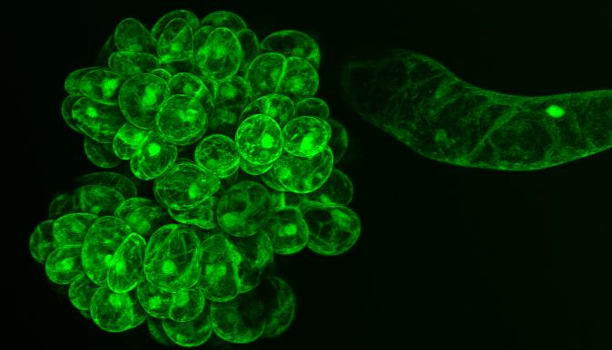
Above second picture from the left is GFP in a-synuclein, the third picture is GFP in moss protoplasts and the forth picture is GFP in transgenic mouse retinas.


More Information:
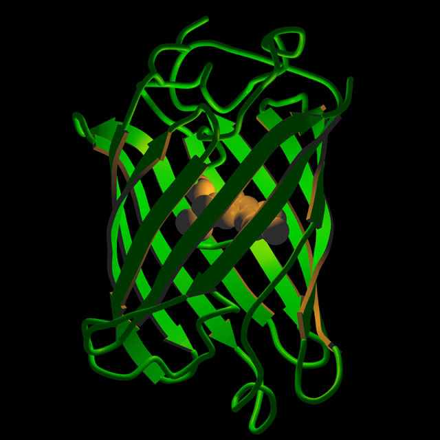
-www-bioc.rice.edu/Bioch/Phillips/Papers/gfpbio.html (Explains the molecular structure of GFP)
-nobelprize.org/nobel_prizes/chemistry/laureates/2008/chemadv08.pdf (Discovery and development of GFP)
-www.sciencedaily.com/releases/2008/10/081008100616.htm (2008 Nobel Prize Article)
-www.callutheran.edu/Academic_Programs/Departments/BioDev/omm/gfp/gfp.htm (Structure of GFP)
-flytrap.med.yale.edu/ (GFP experiment)
-www.rpc.msoe.edu/cbm2/gfp1.htm (What GFP is)
No comments:
Post a Comment